


I. Introduction
Cellulitis is a prevalent bacterial skin infection that can potentially have serious consequences. This infection targets the middle skin layer(dermis) as well as the underlying tissues, occasionally extending to the muscle beneath the skin1).
The infection happens when a break in the skin allows bacteria to enter. There's evidence indicating that the existence of fungal foot infections like tinea pedis and toenail onychomycosis can increase the risk of cellulitis within the lower limb. Moreover, factors such as a history of peripheral vascular disease and the utilization of corticosteroid medications or other immune-suppressing drugs are also recognized as risk factors for the development of cellulitis2,3).
Cellulitis typically resolves within 7 to 10 days with the use of antibiotics4). However, people with fungal foot infections, particularly those who have diabetes, may experience recurrent cellulitis3). When cellulitis is left untreated or if the treatment is ineffective, it can lead to various complications such as lymphangitis, sepsis, osteomyelitis, endocarditis, meningitis, shock, and more5). To address the infection and alleviate cellulitis severity, treatment should consider immune stimulatory activity. This immune support is crucial for promoting tissue repair and regulating inflammation6).
Sodokum, first documented in the Ming Dynasty's 'Uigamseo', has been used in the treatment of rashes. Some studies have shown its antibacterial effects against various skin pathogens, antioxidant properties, and anticancer effects7). Sipjeondaebo-tang, Samul-tang plus Sagunja-tang, has immune stimulatory activity, showing splenocyte proliferation and macrophage proliferation8).
Besides the cases reported by Lee et al.9), Choi et al.10), and others11), there are limited case reports on Korean medical treatment for cellulitis. We confirmed symptom improvement in the patient with cellulitis through enhanced local immunity with the combination of Sodokum and Sipjeondaebo-tang.
This case report describes Korean medical treatment for cellulitis.
II. Objects & Methods
We studied one patient who developed cellulitis after sustaining toe wounds caused by a foot fungus. The patient has been fully informed and has given consent for the use of their information in publishing this paper.
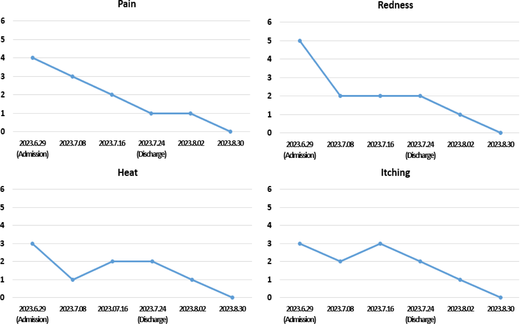
The Visual Analog Scale(VAS) is a measurement tool used to assess subjective experiences, primarily pain, discomfort, or other subjective sensations. The VAS was utilized to evaluate the degree of discomfort perceived by the patient, including pain, redness, heat, and itching. The score ranged from 0 to 10, with 0 indicating no sensation or discomfort and 10 indicating the most severe or intense experience.
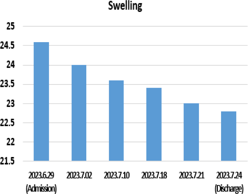
To assess edema, we measured the circumference of the left foot passing through the 太衝(LR3) acupuncture point, which served as the central point of the lesions, in centimeters.
III. Case
-
Patient : ○○○(F/76/Height 153㎝/Body weight 60.3㎏)
-
Treatment period : 2023.6.28.–2023.8.30 (Admission : 2023.6.29–2023.7.24, Outpatient clinic visit dates : 2023.8.2, 2023.8.30)
-
Chief complaint : Pain, redness, heat, itching, and swelling of the left foot
-
Onset : 2023.6.7
-
Past history : Left tinea pedis & unguium
-
Family history : None of specific
-
Social history : None of specific
On June 7, 2023, a 76-year-old female patient experienced sudden pain, redness, heat, and swelling in her left foot, along with a preexisting condition of tinea pedis and unguium. She sought medical attention at the internal medicine department on June 14, 2023, where she was diagnosed with cellulitis. She took cephalosporin antibiotics(Suprax capsule) for 7 days. On June 28, 2023, she began Korean medical treatment for her condition.
The patient took 3 times a day during June 29, 2023 to July 24, 2023. The same medicine was prescribed for 10 days at discharge(Table 1).
We instructed the patient to use Chisun solution(containing Hibisci Cortex(木槿皮), Salicylic acid, etc., produced by Hani External Therapy Association) to relieve symptoms of athlete's foot. Additionally, for redness and heat relief, we recommended the application of Sambaekihwan-go(manufactured by Hani External Therapy Association) to the affected area as needed. During the hospitalization period, a daily wet dressing was applied to the left foot for 20 minutes each day using an external prescription of our hospital(Table 2). Upon discharge, the patient received instructions on wet dressing procedures and continued the regimen at home using the same external prescription for a consecutive 15 days after discharge.
| Herbal name | Scientific name | Dose (g) |
|---|---|---|
| 樺皮 | Betulae Cortex | 10 |
| 梔子 | Gardeniae Fructus | 15 |
| 黃連 | Coptidis Rhizoma | 10 |
| 苦參 | Sophorae Radix | 15 |
| 白鮮皮 | Dictamni Radicis Cortex | 10 |
| Total | 60 |
The acupuncture needles were standardized stainless steel, 0.25×40㎜, and disposable (manufactured by Boryung, a Korean Eastern acupuncture equipment manufacturer). Acupuncture was administered at the affected area of the left foot for 15 minutes once a day during the hospitalization period. The depth of the acupuncture needle insertion was approximately 3-5 millimeters.
At admission, the patient reported throbbing pain, particularly around the left ankle, with a VAS score of 4. Due to this pain, walking for more than 5 minutes without assistance from a monocane was not possible. Redness, heat, and itching sensations were rated as VAS 5, VAS 3, and VAS 3, respectively. Standing or moving exacerbated the redness, eventually causing the left foot to turn black.
Upon discharge, the pain decreased to approximately VAS 1, allowing the patient to walk for around 20 minutes without monocane assistance. Both redness, heat, and itching sensations significantly decreased to VAS 2.
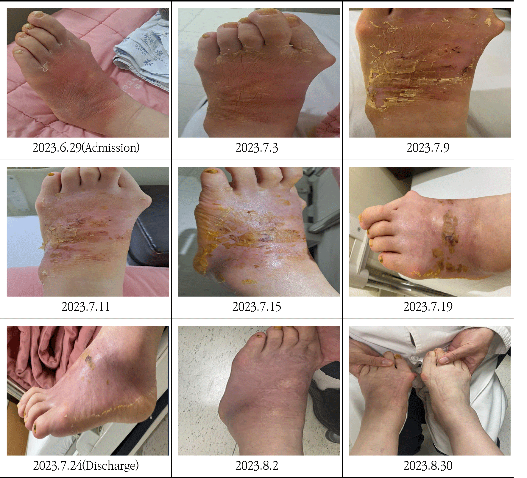
At the outpatient follow-up on August 2, 2023, subjective symptoms, including pain, redness, heat, and itching, reduced to VAS 1. In the final follow-up on August 30, 2023, not only was there no recurrence of symptoms, but all symptoms were almost completely resolved to the point of the patient enjoying hiking(Fig. 1).
It decreased by approximately 10%, from 24.6㎝ at admission to 22.8㎝ at discharge(Fig. 2).
Throughout the hospitalization, the skin exhibited a pattern of epidermal shedding as part of the regeneration process. The lateral malleolus was the site where pain and redness were most severe. Approximately 10 days after discharge, the skin color had further lightened, and in the final outpatient follow-up, it had almost returned to normal compared to the unaffected side(Fig. 3).
In the blood test conducted immediately after admission, the CRP level was elevated at 0.69㎎/㎗, surpassing the normal range of 0-0.50㎎/㎗. However, in the blood test performed five days after admission, the CRP level significantly decreased to 0.16㎎/㎗. Looking at the complete blood cell count results, during the early hospitalization period, the monocyte level slightly exceeded the normal range of 2-9%, but it returned to within the normal range on the third blood test(Table 3).
IV. Discussion
Cellulitis occurs most frequently in the lower extremities and usually unilateral2). Clinically, patients experience redness, swelling, increased temperature, and heightened sensitivity in their skin1). The most recent guideline from The Infectious Disease Society of America(IDSA) on skin and soft tissue infections states that most cases of cellulitis are due to streptococci, often Streptococcus pyogenes, but also groups B, C, or G. S. aureus is also noted to cause cellulitis, especially when associated with furuncles, carbuncles, or abscesses4).
Unfortunately, there is no definitive or reliable diagnostic method for cellulitis. Routine biochemical and hematological blood tests, as well as blood cultures, lack specificity in confirming the diagnosis13). Recently, there has been a recognition that change in peripheral blood leukocyte ratios serves as a simple, rapid, and novel inflammation parameter for several diseases. Specifically, blood monocytes offer insight into the systemic immune response, from production to tissue recruitment, reflecting the impact of infection on the host12,14).
Skin infections are the most common cause of lymphangitis. Lymphangitis can occur in the setting of normal lymphatic channels with acute infection, damaged lymphatic channels, or anatomic abnormalities. Acute lymphangitis may occur in the setting of skin abrasion with infection at a distal site, such as interdigital dermatophyte infection or cellulitis or erysipelas of the lower leg. Lymphatic damage and anatomic abnormalities can result in tissue protein and fluid accumulation, leading to nonpitting lymphedema with induration and predisposing to invasion of microorganisms. Particularly, when standing, the effects of gravity can contribute to heightened fluid buildup and impaired blood circulation around the inflamed area, potentially exacerbating pain and redness15).
To evaluate the results, we checked not only whether symptoms improved and whether there was a recurrence but also inflammatory markers through blood tests and observed lesion changes in photographs. The patient, experiencing heightened pain upon standing and moving due to infection extending to the underlying tissues, was unable to walk for more than 5 minutes without assistance from a monocane upon admission. If standing or moving persisted beyond 10 seconds, pain and redness intensified, eventually leading to the skin of the left foot turning black. Additionally, the circumference of the affected foot was 24.6㎝. However, at the time of discharge, the patient could walk for approximately 20 minutes without monocane assistance, and the circumference had reduced to 22.8㎝. We also confirmed that the patient exhibited a decrease in both CRP and monocyte levels after hospitalization. At the one-month follow-up after discharge, the patient reported no issues with independent walking and had sufficiently recovered to enjoy hiking.
This case signifies a significant outcome as it validates improvement in both subjective and objective symptoms, including edema circumference and hematological measures, following 26 days of hospitalization treatment. During the two outpatient follow-up visits, there was no recurrence of symptoms, and all symptoms were almost completely resolved. This is a meaningful result as it indicates the disappearance of subjective symptoms and no recurrence after only Korean medical treatment, which lasted for about one month.
In our study, 'Sodokum plus Sipjeondaebo- tang' was used as herbal medicine treatment based on the principle of 'strengthening the body's resistance to eliminate pathogenic factors(扶正袪邪)'. When inflammation arises in the body, the immune system expends significant energy combating the infectious agent, leading to a decline in overall health and vitality. Consequently, the treatment of infectious diseases should not only focus on eliminating the pathogen but also consider the restoration of the patient's energy levels6).
Arctii Semen(牛蒡子) and Peucedani Radix(防風), the components of Sodokum, are known for dispelling wind and heat. Additionally, Arctii Semen and Schizonepetae Spica(荊芥) detoxify and clear up rashes to prevent complications7). Sipjeondaebo-tang is a commonly prescribed herbal formula in Korea, Japan, and China. It effectively addresses both qi and blood deficiency syndromes by harmonizing Yin and Yang. Widely employed in the treatment of chronic illnesses, Sipjeondaebo-tang restores physiological function and boosts immunity. Previous studies highlight diverse biological properties of Sipjeondaebo-tang, including anti-cancer, anti-inflammatory, gastric protective, and immune cell activation effects8).
In this case, the patient exhibited no significant issues with sleep or appetite but experienced reduced energy due to infectious diseases, presenting with a pale tongue and thin white coating, along with a thready, soft, deep, and weak pulse. The diagnosis indicated both qi and blood deficiency syndromes resulting from the pathogen, leading to the prescription of this herbal medicine.
Examining the external application treatment utilized in this study, the Chisun solution, featuring Hibisci Cortex as its primary component, is renowned for its outstanding efficacy in athlete's foot treatment with excellent anti-ringworm and anti-inflammatory effects16). Sambaekihwan-go is comprised of powdered Angelicae Dahuricae Radix(白芷), Bletillae Rhizoma(白芨), Alunitum Siccus(枯白礬), Sulphur(硫黃), and Phellodendri Cortex(黃柏). It is known for its heat-clearing properties and itching reduction, proving effective against various bacterial and fungal skin diseases17). Wet dressing contributes to a cooling effect by absorbing the skin's heat, thereby aiding in the reduction of inflammation. Simultaneously, it alleviates itching sensations stemming from the shedding epidermis during the wound recovery process17,18).
We conducted acupuncture treatment on affected sites for the anti-inflammatory mechanism through microtrauma. The acupuncture stimulus induces local microtrauma, triggering the activation of neuro-immune reflexes. These include mast cell involvement and the release of vasoactive substances such as cGRP, histamine, SP, adenosine, prostaglandin, bradykinin, etc. This leads to vasodilation in small vessels, increased blood flow, and the initiation of local inflammatory and anti- inflammatory responses19). In addition, carbon-arc-light irradiation treatment was performed to aid in wound regeneration, metabolism, and swelling absorption during acupuncture treatment20).
This case highlights the potential effectiveness of Korean medicine treatment in alleviating cellulitis, suggesting positive outcomes when employing Korean medicine for cellulitis symptoms.
Research on cellulitis within the realm of Korean medicine is not only confined to case reports but is also notably scarce in quantity. Therefore, further research is imperative. This case report holds significance as it demonstrated improvements solely through Korean medicine interventions, including herbal medicine, external applications, and acupuncture.
However, deriving broad conclusions about the effectiveness of Korean medicine treatment for cellulitis from this single case is challenging. Anticipating more cases in the future, we hope to see elevated research, including experimental considerations and randomized controlled trials(RCTs), to enhance the level of evidence for treatment.