


I. Introduction
Tinnitus, the perception of sound without an external source, affects millions worldwide, yet its underlying mechanisms remain unclear1). Research indicates that tinnitus may initially arise from abnormalities in the cochlear duct, but persistent neural changes in the central auditory system are likely responsible for sustaining the condition2,3). The cochlear duct is sensitive to changes in blood flow, and circulation disorders can lead to dysfunction of the cochlea4). According to Jastreboff's neurophysiological model, when tinnitus becomes severe or chronic, the autonomic nervous system and limbic system play crucial roles in maintaining and influencing the condition5,6).
Effective treatment for tinnitus requires a comprehensive approach due to its involvement with the circulatory, limbic, and autonomic nervous systems. Current clinical treatments include medications, cochlear implants, hearing aids, cognitive behavioral therapy (CBT), tinnitus retraining therapy (TRT), and biofeedback7). However, these often provide limited relief for chronic or severe cases. Acupuncture and moxibustion, traditional Asian medicine treatments, have been demonstrated to alleviate tinnitus symptoms8,9). These treatments are believed to increase local blood flow, modulate the amygdala-limbic system connection, improve olivocochlear nucleus plasticity, and enhance nervous system function10).
In recent years, the acceleration photoplethysmogram (APG) has emerged as a non-invasive tool for assessing cardiovascular function and blood flow dynamics11,12). The pulse wave is measured by the change in the amount of light absorbed by hemoglobin, which generally reflects changes in blood volume. The APG, derived from differentiating the pulse wave twice, simplifies the identification of pulse wave peaks and their timing, allowing for detailed analysis of pulse waveforms11,12). Given the potential association between tinnitus and vascular abnormalities or altered blood flow in the auditory system, the APG could serve as a valuable biomarker for evaluating the effects of acupuncture and moxibustion on tinnitus patients.
Although the number of individual studies exploring acupuncture or moxibustion as treatments for tinnitus is increasing, there is still a lack of research investigating the effects of these therapies using APG. This study aims to examine the effects of acupuncture and moxibustion in tinnitus patients by measuring changes in the APG before and after treatment. By analyzing the APG, we seek to better understand the hemodynamic effects of these interventions and their potential role in managing tinnitus.
II. Methods
A retrospective chart review was conducted on patients with tinnitus who visited Dongguk University Ilsan Oriental Hospital between April 2024 and July 2024. The research adhered to the ethical guidelines outlined in the Declaration of Helsinki for research involving human subjects. This study was approved by the Institutional Review Board of Dongguk University Ilsan Oriental Hospital (approval number: DUIOH 2024-06-006-001). As this was a retrospective chart review, informed consent was waived. All patient information was obtained from existing medical records.
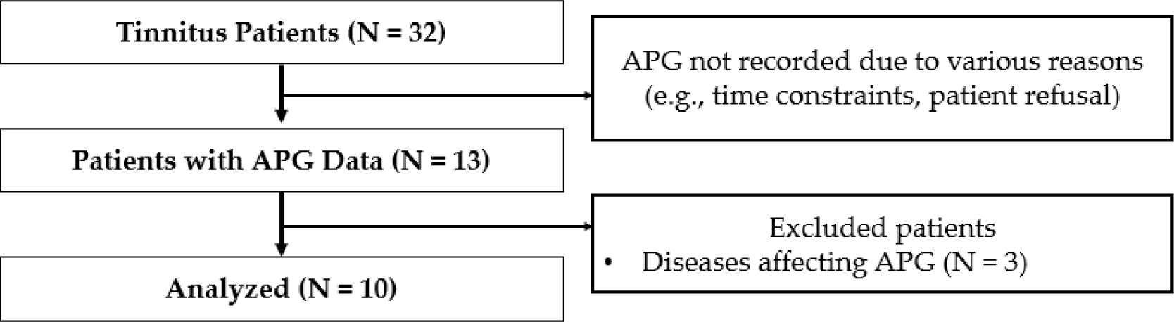
This study involved a review of medical records for patients with subjective non-pulsatile tinnitus who sought care at the Ophthalmology, Otolaryngology, and Dermatology outpatient department (OPD) at Dongguk University Ilsan Oriental Medicine Hospital in Goyang, Republic of Korea, between April and July 2024. Patients with available APG data were included in the study. Information collected from the electronic medical records included patient demographics, comprehensive medical history, medications, tinnitus details, APG data, treatment specifics, and treatment outcomes (Fig. 1).
The exclusion criteria were;
-
(1) Presence of diseases known to affect APG (cardiovascular, endocrine, autoimmune, respiratory, neurologic, and psychiatric disorders, including alcoholism and polytoxemia)
-
(2) A history of smoking
-
(3) Use of vasoconstrictors, beta-blockers, anticholinergics, antidepressants, or other central nervous system-active medications within the past four weeks
-
(4) Pregnancy
APG measurements were conducted three times during a single OP visit: before treatment, after acupuncture, and after moxibustion. After an initial consultation, participants removed any metal objects and lay down in comfortable beds in a quiet, temperature-controlled room. Patients relaxed for 10 minutes before the initial measurement, and subsequent measurements were taken immediately after each treatment. All APG measurements were taken using the uBioMacpa device (Biosense Creative Inc., Seoul, Republic of Korea).
The acupuncture needles were standardized, stainless steel, 0.25×40㎜, and disposable (manufactured by Boryung, a Korean Eastern acupuncture equipment manufacturer). Acupuncture was applied to the following points: Baekhoe (GV20), Yepung (TE17), Imun (TE21), Pungji (GB20), and Cheonggung (SI19) on the affected side, as well as Duyu (ST8) and Hapgok (LI4)11)on both sides. The depth of needle insertion was approximately 3-5㎜. Needle manipulations were performed to achieve the de qi sensation. The needles were left in place for 15 minutes without additional manipulation.
After acupuncture, the same practitioner applied non-contact moxibustion to the SI19 point on the affected side. A moxa roll made from dried mugwort leaves (Dongbang Medical, Dongbang Moxa) was held about 2–3 cm above the skin. The procedure was repeated three times for a total duration of 10 minutes. Patients experienced a warm sensation, similar to the ‘Deqi’ sensation in acupuncture, before skin redness occurred.
The following descriptive variables were obtained from participants' electronic medical records.
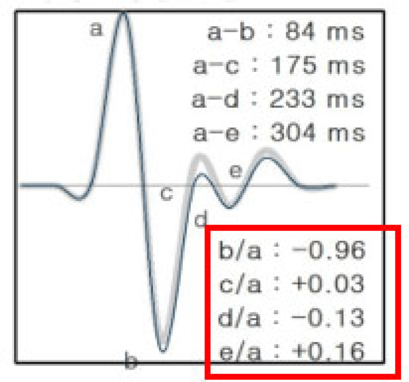
The APG waveform consists of four systolic waves and one diastolic wave, specifically the a-wave (early systolic positive wave), b-wave (early systolic negative wave), c-wave (late systolic re-increasing wave), d-wave (late systolic re-decreasing wave), and e-wave (early diastolic positive wave). The height of each wave was measured from the baseline, with positive values above the baseline and negative values below it. The ratios of the height of each wave relative to the a-wave (b/a, c/a, d/a, and e/a) are typically used in wave analysis12)(Fig. 2).
Statistical analysis was conducted using Python 3.13, with significance set at P<0.05. Continuous variables were presented as means with standard deviations (SD) or standard errors (SE), while categorical variables were summarized as counts and percentages. Linear Mixed-Effects Models (LMMs) were used to assess treatment effects on APG parameters, with treatment as a fixed effect and patients as random effects. The normality of residuals from the LMMs was verified with the Shapiro-Wilk test. To analyze changes in APG values for each patient across multiple OP visits, pre- and post-treatment differences were also tested for normality. Depending on the results, either a paired t-test or Wilcoxon signed-rank test was applied.
III. Results
The clinical characteristics of the tinnitus patients (N = 10) are summarized as follows: Sixty percent of the patients (6 individuals) were male, with a mean age of 41.8 years ± 18.4 (SD). The average duration since tinnitus onset was 12.1 months ± 11.5 (SD). Regarding the tinnitus site, 80% of patients (8 individuals) had unilateral tinnitus, while 20% (2 individuals) had bilateral tinnitus. The mean number of outpatient visits was 6.9 ± 3.9 (SD) (Table 1).
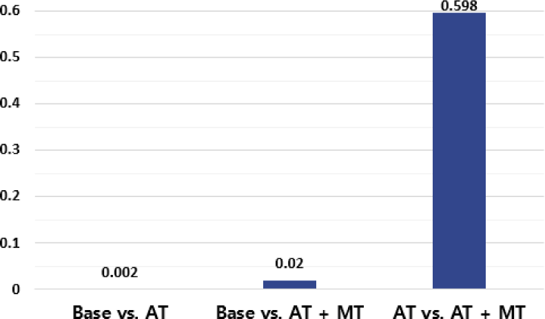
This study applied a linear mixed-effects model to account for individual patient variability when analyzing treatment effects across three groups: Base (before treatment), AT (after acupuncture), and AT + MT (after acupuncture and moxibustion). The mean values and standard deviations for the ‘b/a’, ‘c/a’, ‘d/a’, and ‘e/a’ ratios across these conditions are presented (Table 2). The ‘e/a’ ratio consistently showed positive values. Significant differences in the ‘c/a’ ratio between Base vs. AT (Effect = 0.053, P= 0.002) and Base vs. AT + MT (Effect = 0.043, P= 0.020) are highlighted (Table 3, Fig. 3). The ‘b/a’ ratio did not show significant changes, with P values ranging from 0.425 to 0.739. No significant differences in the ‘d/a’ and ‘e/a’ ratios across any comparisons are indicated (Table 4). Additionally, the comparison between AT and AT + MT showed no significant differences across all indicators, suggesting that the addition of moxibustion did not provide further improvement. Overall, group variability was generally low across all comparisons, indicating consistent effects within each group, with some indicators, such as ‘e/a’, showing minimal to no variability.
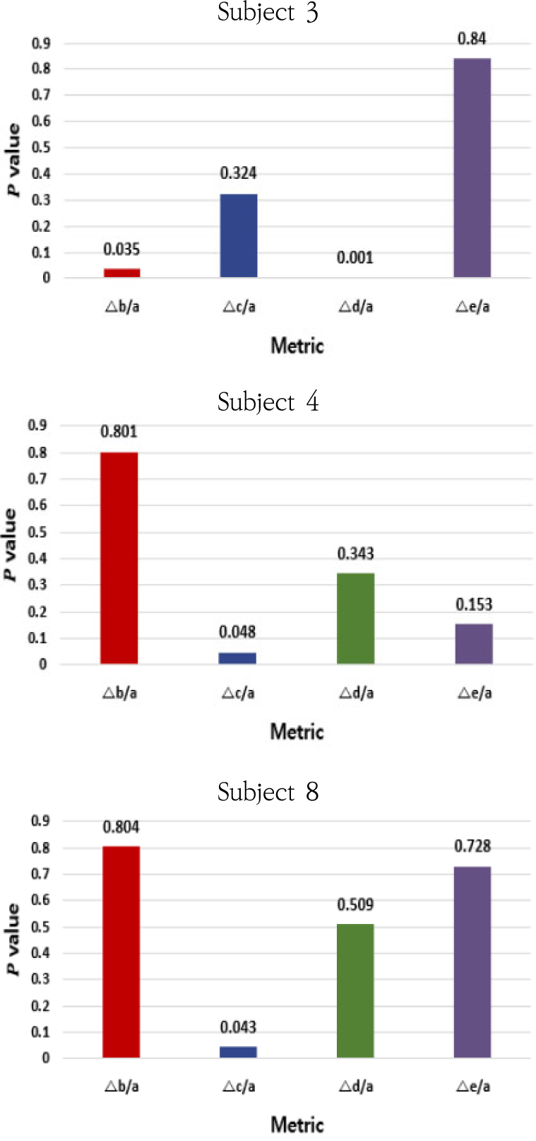
P-values for the differences in APG metrics (△b/a, △c/a, △d/a, △e/a) among eight subjects who had more than three OP visits are presented (Table 5). As a result of testing whether the mean of these differences is statistically significantly different from zero, significant changes were observed in Subject 3, specifically in the △b/a (P= 0.035) and △d/a (P = 0.001) metrics. Additionally, Subject 4 showed a significant change in the △c/a metric (P= 0.048), and Subject 8 also exhibited significance in △c/a (P= 0.043). No significant differences were detected in the other subjects (P> 0.05) (Fig. 4).
IV. Discussion
This study investigated the hemodynamic effects of acupuncture and moxibustion in tinnitus patients by analyzing changes in APG parameters, with a focus on the b/a, c/a, d/a, and e/a ratios. Our findings revealed a significant increase in the c/a ratio following acupuncture, while the other ratios remained unchanged.
While the underlying mechanisms of tinnitus are complex and multifactorial, research suggests that it may originate from abnormalities in the cochlear duct2-4). The inner ear, particularly its hair cells, is highly sensitive to fluctuations in blood flow, and its function can be impaired by hypoxic conditions resulting from microcirculatory disturbances4). Studies have identified associations between tinnitus and various systemic conditions, such as cardiovascular disease, dyslipidemia, and diabetes14-16). Pharmacological interventions for tinnitus often focus on improving blood flow through agents such as vasodilators, atorvastatin, and ginkgo biloba extract17). Acupuncture and moxibustion have been widely employed as alternative therapeutic approaches for tinnitus, due to their ability to improve blood circulation18,19).
Techniques like functional near-infrared spectroscopy (fNIRS), functional magnetic resonance imaging (fMRI), and Technetium-99m Ethyl Cysteinate Dimer Single Photon Emission Computed Tomography (Tc-ECD SPECT) have been used to investigate cortical blood flow in tinnitus patients20-23). However, these methods are costly, time-intensive, and require specialized facilities, limiting their utility for routine or large-scale studies
In contrast, PPG offers several advantages for hemodynamic assessment12). PPG sensors are non-invasive, portable, and cost-effective tools that can be easily applied to any superficial vessel24). They allow for real-time monitoring of peripheral blood flow and cardiovascular function in both clinical and non-clinical environments24,25). The second derivative of PPG, APG, further enhances cardiovascular analysis by providing more detailed insights than the first derivative24-26). Considering that tinnitus is associated with systemic circulatory disturbances, incorporating APG into research could enhance our understanding of the hemodynamic effects of acupuncture and moxibustion in tinnitus patients by complementing existing cerebral blood flow studies.
The APG waveform includes four systolic waves and one diastolic wave, with the b/a, c/a, d/a, and e/a ratios serving as key indicators of arterial stiffness and cardiovascular health12,13,27-29). The c/a ratio, which reflects the relationship between the late systolic reincreasing wave (c-wave) and the early systolic positive wave (a-wave), is closely associated with arterial elasticity and typically decreases with age as arterial stiffness increases12,27-29). The significant rise in the c/a ratio after acupuncture suggests a decrease in arterial stiffness. This result is consistent with a previous study showing a notable reduction in the APG-d/a index following acupuncture at the Pericardium 6 (PC6) acupoint, indicating a decrease in left ventricular afterload30).
Previous research has shown that acupuncture can enhance arterial flexibility by modulating autonomic nervous system activity, particularly by increasing parasympathetic tone and reducing sympathetic outflow31). This improvement in the c/a ratio may indicate an enhanced vascular environment around the auditory system, reducing neural hyperactivity and helping to mitigate the neural mechanisms that sustain tinnitus8-10). These findings suggest that acupuncture could be a particularly beneficial treatment option for individuals with tinnitus related to autonomic dysregulation or vascular abnormalities8-9).
Conversely, the b/a, d/a, and e/a ratios did not show significant changes following acupuncture or moxibustion. The b/a ratio, which is associated with increased arterial stiffness and typically rises with age12,13), might not have been significantly affected because of the short duration of the intervention or the specific characteristics of the patient population, such as their baseline vascular health. Similarly, the d/a ratio, which reflects both decreased arterial stiffness and left ventricular afterload12,27), might require more intensive or prolonged treatment to exhibit meaningful changes. The e/a ratio, indicative of diastolic function and the prominence of the dicrotic notch12), also remained stable, suggesting that acupuncture primarily influenced systolic rather than diastolic vascular dynamics.
The addition of moxibustion did not lead to significant changes in any of the APG ratios compared to acupuncture alone. While moxibustion is intended to warm the meridians and promote qi and blood circulation19), the lack of further improvements suggests that acupuncture may already achieve the maximum vascular response, or moxibustion's impact on vessel compliance, as measured by APG, may be less direct than that of acupuncture.
In addition to the group-level findings, individual patient analysis revealed significant changes in APG metrics for specific subjects. For each patient, the differences in indicators (b/a, c/a, d/a, e/a) before and after treatment were calculated. In our study, we tested whether the mean of these differences was statistically significant from zero. Specifically, significant alterations were observed in the b/a and d/a ratios for Subject 3, and in the c/a ratio for Subjects 4 and 8. These individual variations suggest that the hemodynamic response to acupuncture may vary among patients, possibly due to differences in vascular health, tinnitus severity, or individual physiological responses to treatment. This emphasizes the need for a personalized approach to acupuncture therapy for tinnitus, where the selection and combination of acupoints are tailored to individual patients' specific needs to maximize therapeutic benefits.
This study has several limitations. The small sample size of 10 participants restricts the generalizability of the findings. The retrospective design, which relies on existing medical records, introduces the risk of information bias due to potential inaccuracies or incompleteness in the data. Furthermore, the four-month study period may not have captured the full range of patient variability or the long-term effects of acupuncture and moxibustion. Additionally, the study primarily focused on the immediate effects of the treatments, limiting our understanding of their long-term impact on the hemodynamic status of tinnitus patients.
Compared to other cardiovascular monitoring methods, such as electrocardiography (ECG), PPG and APG have drawbacks. Unlike ECG, which directly measures the heart's electrical signals with high accuracy, PPG and APG rely on optical methods to infer blood volume changes, making them more susceptible to distortion and less reliable for precise cardiovascular assessments under certain conditions12,24). The most significant weakness of PPG sensors is their vulnerability to motion artifacts24). Additionally, ambient light conditions can influence PPG measurements, requiring careful positioning of LEDs and photodetectors, as well as ensuring optimal sensor-skin contact24). Skin color can also impact results32). These limitations may explain why, in our study, certain APG ratios like b/a, d/a, and e/a did not exhibit significant changes post-treatment. Detecting subtle hemodynamic changes in these ratios may require longer observation periods or more controlled conditions to achieve accurate results.
Future research should employ larger, randomized controlled trials to validate these findings and investigate the long-term effects of acupuncture and moxibustion on APG parameters and tinnitus symptoms. To achieve this, studies should extend treatment durations, increase sample sizes, and incorporate additional hemodynamic biomarkers. Moreover, they should examine whether specific patient characteristics, such as baseline cardiovascular health or the duration and severity of tinnitus, are predictive of significant hemodynamic changes in response to acupuncture.
In conclusion, this study demonstrates that acupuncture may induce beneficial hemodynamic changes in tinnitus patients, particularly through the reduction of arterial stiffness, as indicated by significant alterations in the c/a ratio observed both at the group level and in specific individual cases. However, the observed variability in individual responses underscores the necessity for personalized treatment approaches and further research to optimize acupuncture and moxibustion protocols for the effective management of this condition. Comprehensive investigations into tinnitus biomarkers are needed to fully understand the mechanisms underlying these effects and to refine therapeutic strategies aimed at maximizing the benefits of these treatments.
V. Conclusion
In tinnitus patients, acupuncture or combined acupuncture and moxibustion were shown to induce hemodynamic changes, particularly reflected in significant alterations in the c/a ratio, indicating a reduction in arterial stiffness. Additionally, individual patient analyses revealed significant changes in specific APG indices for some patients. The variability in these individual responses highlights the importance of personalized treatment approaches and the need for further research to optimize acupuncture and moxibustion protocols for effective tinnitus management.